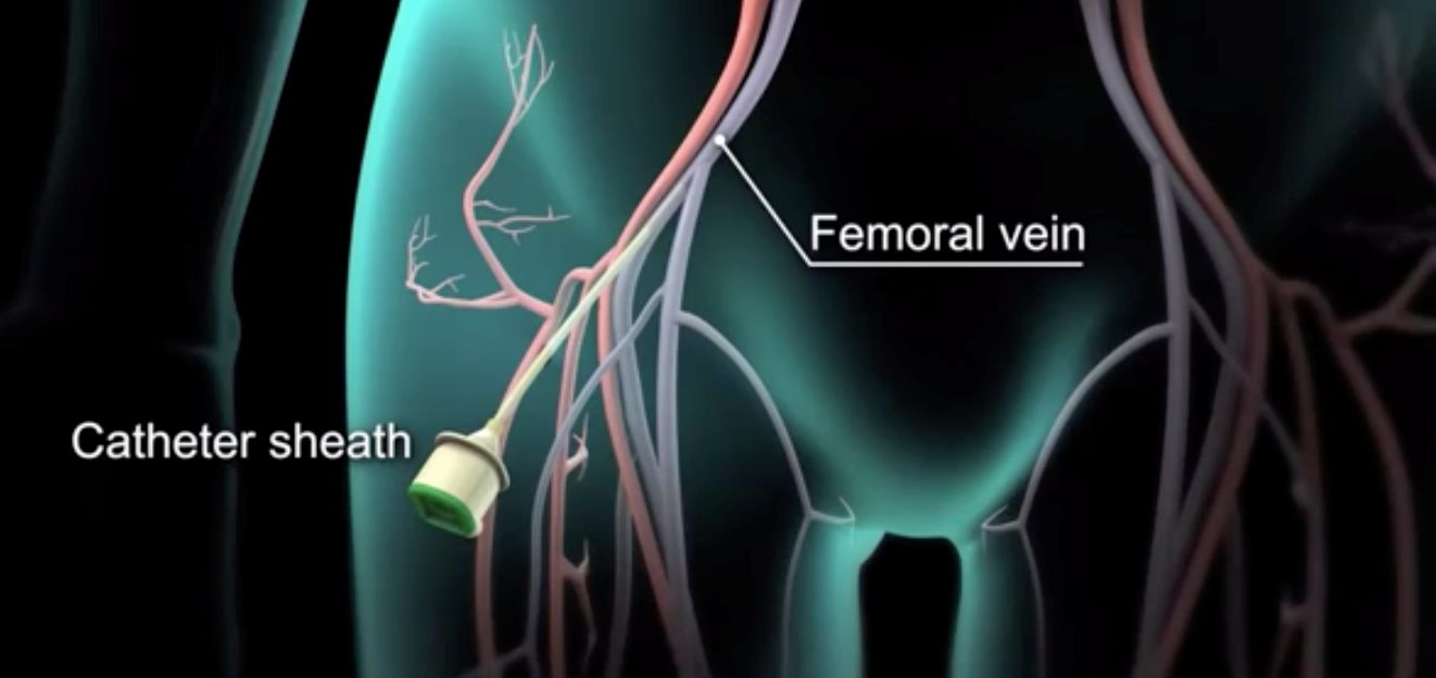
The procedure requires placement of a sheath inside one of the blood vessels of the leg (commonly femoral vein). Through this femoral vein, some tubes (catheters) are introduced inside the heart which detect the electrical problem. This is called electrophysiological study. It is done with local numbing agent so the patient will not feel any pain.

The tubes can be inserted inside upper chamber(s) of the heart or lower chamber(s) of the heart, depending upon the type of arrhythmia that the patient has. After this mapping is performed to find out the source electrical problem in the heart. Two common types of mapping systems are – two-dimensional and three-dimensional mapping system.
Once the electrical problem is identified, it is treated with application of local energy in a procedure called as ablation. Common energy used is electrical energy so the ablation is called radiofrequency ablation. This energy application is inside the heart so the patient may feel it but it will not cause pain.